top of page

PHOTO & VIDEO GALLERY
Here is a collection of photos and videos of our experiments and days in the lab.
Photos
 The micropipette we will use for various experiments in this lab |  Our template DNA and primers being kept on ice |
|---|---|
 Weighing out water to balance the centrifuge |  The collective vial is put into the centrifuge with a vial of water of equal mass opposite to balance |
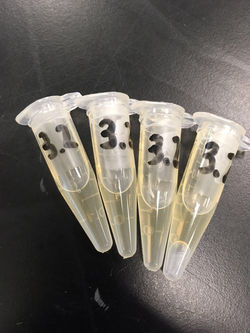
 Our group's vial |  Patrick bringing up our vial on ice to the thermal cycler |
 The thermal cycler machine will run the samples through the process of PCR |  Putting our vial in the thermal cycler |
 The various group's vials in the thermal cycler |  The results of the gel electrophoresis |
 All of the necessary materials for the yeast transformation |  Sterile water for the yeast transformation |
 Salmon sperm for the yeast transformation |  A beaker for the supernatant waste |
 Our vial where we mixed all the components for the yeast transformation |  The vortex machine used to mix up the vials |
 The centrifuge machine used in the yeast transformation |  The 96-well plate we would use for a practice serial dilution |
 Putting the 96-well plate into the plate reader (spectrophotometer) | 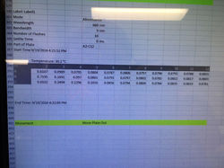 The data from our practice spectrophotometer run |
 The graphed results of our practice run with the spectrophotometer | 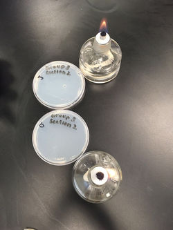 Practicing our sterile technique with ethanol burners |
 Pipetting our yeast onto agar plates |  Pipetting our yeast onto agar plates close to ethanol burner to avoid contamination |
 The YPD media we made |  Our YPD jar in the autoclave machine |
 Our liquid YPD tubes |  The Fluorescence Activated Cell Sorting (FACS) machine |
 A description of each of our agar plates |  First set of pictures of colonies for WT yeast in normal conditions |
 First set of pictures of colonies for mutant yeast in normal conditions |  First set of pictures of colonies for WT yeast on caffeine agar |
 First set of pictures of colonies for mutant yeast on caffeine agar |  First set of pictures of colonies for WT yeast on bleomycin agar |
 First set of pictures of colonies for mutant yeast on bleomycin agar |  The 96-well plate with our YPD and yeast for growth curve analysis |
 Growth curves for WT strain |  Growth curves from mutant strain |
 Our 3D model from Tinkercad |  Our printed wax paper |
 Melting the wax paper to create a hydrophobic barrier |  Covering the back of our wax paper with vaseline |
 Pipetting agar onto the white squares of our wax paper |  Pipetting agar onto the white squares of our wax paper |
 The contamination on our first wax paper trial |  Putting nail polish on the wax paper for second trial |
 Our sterile setup for the wax print experiment |  The updated design of our 3D model |
 First successful trial of wax paper experiment |  First successful trial of wax paper experiment |
 First successful trial of wax paper experiment |  First successful trial of wax paper experiment |
 Wax paper experiment with optimal square only |  Optimal square trial results |
 Optimal square trial results |  Table of agar ratios used on different sized squares |
 Our model in the dried PDMS |  3D print |
 2nd 3D print with structural supports |  Covering our 3D model with PDMS |
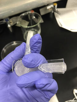 Our model covered in PDMS |  Presenting our poster at the HHMI symposium |
 Suspending our model with the PDMS in NaOH |  The NaOH used to melt away our model |
Videos
This is a video we made explaining homologous recombination repair, polymerase chain reaction and gel electrophoresis.
bottom of page